Scientists’ Engineered Protein Slows Lung Cancer Growth
By: Stephen Fontenot | Oct. 20, 2021
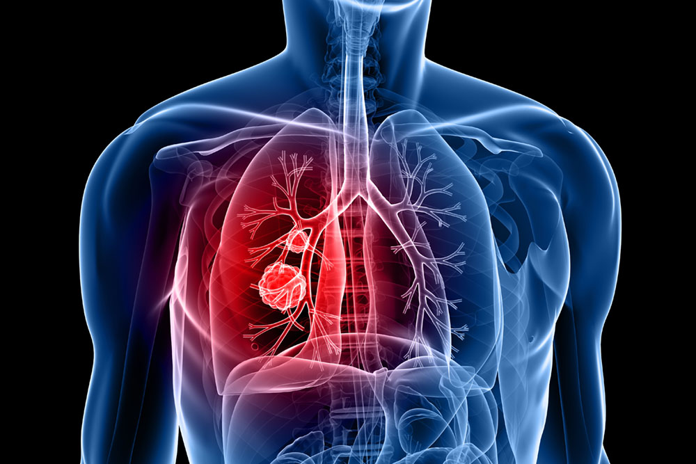
Researchers from The University of Texas at Dallas’ School of Natural Sciences and Mathematics have refined a method they developed to change the environment surrounding lung cancer cells so that chemotherapy or radiation treatment can more easily destroy them.
In a study published online Sept. 22 in Molecular Cancer Therapeutics, scientists show in an animal model that denying tumors access to heme — an iron-binding molecule that is a component of hemoglobin — can stifle lung tumor growth without side effects to other essential biological processes.
Dr. Li Zhang, professor of biological sciences and the Cecil H. and Ida Green Distinguished Chair in Systems Biology Science, is corresponding author of the study. Her research demonstrates that heme-sequestering proteins (HeSPs) designed by her research group slow the growth of non-small cell lung cancer (NSCLC) cells in mice.
“We’re establishing for the first time that this group of therapeutic agents — these engineered proteins — can directly extract heme from hemoglobin,” Zhang said. “Previous demonstrations of this have provided indirect evidence. What we’ve done is to show heme being removed from hemoglobin in a very easily observed manner.”
The ability to remove heme from a tumor environment is important: In an article published in Cancer Research in 2019, Zhang’s team established a link between the presence of heme and lung tumor expansion. In that earlier study, Tianyuan Wang MS’16, PhD’21 — now a postdoctoral researcher at UT Southwestern Medical Center — created heme-sequestering peptides by reengineering hemophores, which are proteins found in bacteria. Hemophores are released by bacterial cells to scavenge a host’s hemoproteins for heme, which carries the iron that bacteria need to survive.

“We’re establishing for the first time that this group of therapeutic agents — these engineered proteins — can directly extract heme from hemoglobin. Previous demonstrations of this have provided indirect evidence. What we’ve done is to show heme being removed from hemoglobin in a very easily observed manner.”
Dr. Li Zhang, the Cecil H. and Ida Green Distinguished Chair in Systems Biology Science in the School of Natural Sciences and Mathematics
Wang, who is a lead author of the Molecular Cancer Therapeutics study, said that the protein design implemented in Zhang’s lab remains unique.
“The idea of using a bacterial hemophore to sequester heme from tumor cells is Dr. Zhang’s original idea,” she said. “No one else has published an idea like that.”
Adnin Ashrafi MS’15, co-lead author of the new study and a molecular and cell biology doctoral student at UT Dallas, said that although the scientists’ heme-sequestering proteins are administered intravenously and, thus, spread throughout the body, no negative side effects have been observed.
“Mice treated with HeSPs did not have significant changes in liver cell ATP [adenosine triphosphate] levels, red blood cell counts or hemoglobin levels,” Ashrafi said. “Because heme synthesis in red blood cells and liver cells is very high, these cells are not affected when heme in an external source — such as a tumor — is depleted.”
Lessening the Growth Level
The researchers used imaging, immunohistochemistry, Western blotting and functional assays to examine human NSCLC cells grafted onto mice. The results showed that heme-sequestering proteins act by affecting oxygen levels and blood-vessel growth in tumor environments.
“Reducing heme suppresses tumors both by limiting tumor hypoxia and normalizing tumor vasculature,” Zhang said. “Multiple mechanisms are at work to restrict the tumors’ growth.”
Wang said tumor hypoxia — oxygen deprivation — aids tumor development by enabling the tumor cells with the most aggressive properties to survive and dominate, while less aggressive tumor cells die.
“Additionally, under hypoxia, tumor cells will generate more blood vessels that are dysfunctional so they cannot deliver a drug effectively to tumors,” Wang said. “Radiotherapy also does not work well in these cases.”


Leaky blood vessels provide cancer cells with excess heme that fuels a tumor’s spread, Zhang said. Employing HeSPs to remove heme helps return blood-vessel growth to normal.
“Normalizing tumor vasculature is very important,” Zhang explained. “In the lung tumor’s environment, the blood vessels are very dense and leaky, which gives tumors abnormally high access to heme. Our protein normalized the blood vessels there — there are fewer blood vessels, and they are structurally intact. The tumor then cannot be as aggressive.”
Zhang emphasized that HeSPs are not a stand-alone treatment; when employed, their deceleration of tumor proliferation, diminished tumor hypoxia and normalized tumor vasculature would improve the chances of radiation, chemotherapy or immunotherapy destroying the tumors.
“Beyond slowing tumor growth, when vasculature is normalized, drugs can be delivered intravenously to the tumors more efficiently,” she said. “Radiation therapy can work better because normalized vasculature prevents tumor hypoxia, which makes tumors more resistant to treatment. In multiple ways, our method normalizes a tumor’s environment.”
Targeting Other Tumors
The researchers also tested the HeSPs on a second type of lung cancer cell, as well as in a breast cancer model. The effect was the same — heme uptake was lowered, and tumor proliferation dropped significantly.
New Dimensions
Learn how to contribute to the New Dimensions: The Campaign for UT Dallas, which aims to enhance lives through transformative research at UTD.
For Zhang and her team, the next steps include testing the combination of heme-sequestering proteins with chemotherapy and radiation therapy in mice. That work will be done in collaboration with UT Southwestern. They will continue to broaden their work to more kinds of cancers, such as kidney cancer, and on drug-resistant tumors.
“We believe we’ve shown that heme sequestration can be a powerful strategy for suppressing lung tumors, and I think that this will apply to drug-resistant tumors as well,” Zhang said.
Additional authors of the paper include molecular and cell biology doctoral students Narges Salamat MS’18, Parinaz Sadat Alemi MS’21 and Eranda Berisha MS’21; biology senior Parsa Modareszadeh; Purna Chaitanya Konduri MS’14, PhD’20, an associate scientist at Austin-based DNA testing company Natera; Sanchareeka Dey PhD’21, a staff scientist at the Retina Foundation of the Southwest; and Poorva Ghosh MS’16, PhD’20, a postdoctoral fellow at UT Southwestern.
This work was supported by a Cancer Prevention and Research Institute of Texas grant.
Media Contact:
Stephen Fontenot, UT Dallas, 972-883-4405, stephen.fontenot@utdallas.edu, or the Office of Media Relations, UT Dallas, (972) 883-2155, newscenter@utdallas.edu.


